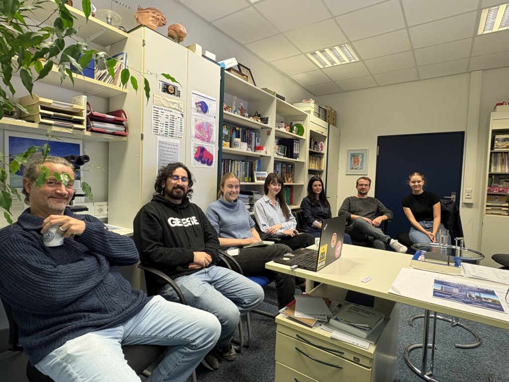
At their lab shadowing meeting on October 14, 2026, projects A02 and A05 discussed the latest results from Kristian Franze’s group. Using the Xenopus model, the group is investigating how a knockdown of SLC35A2 affects the mechanics of the developing brain.
- Sebastián Vásquez presented preliminary evidence that SLC35A2 knockdown (KD) brain tissue becomes stiffer and that cell–cell adhesion forces increase markedly (tether-pulling assay). At the same time, wild-type optic tract axons no longer grow in a targeted manner through KD tissue. This is consistent with the known role of tissue stiffness as a guidance cue.
- Sarah Fritsche is investigating glycosylation in her new project, in particular galactose-dependent polysialylation. To this end, she has set up staining approaches that visualize galactose and various glycan structures (including sialic acids, GlcNAc, and fucose) in tissue.
Taken together, these results and approaches led to the working hypothesis that SLC35A2 loss causes a lack of UDP-galactose in the Golgi. This would alter the glycosylation of adhesion molecules (e.g., N-cadherin, possibly also NCAM1 or neuroligins), thereby stabilizing cell–cell adhesion and increasing tissue stiffness. The participants also agreed on how human tissue samples can be provided for AFM measurements in the future.
Ingmar Blümcke, A02