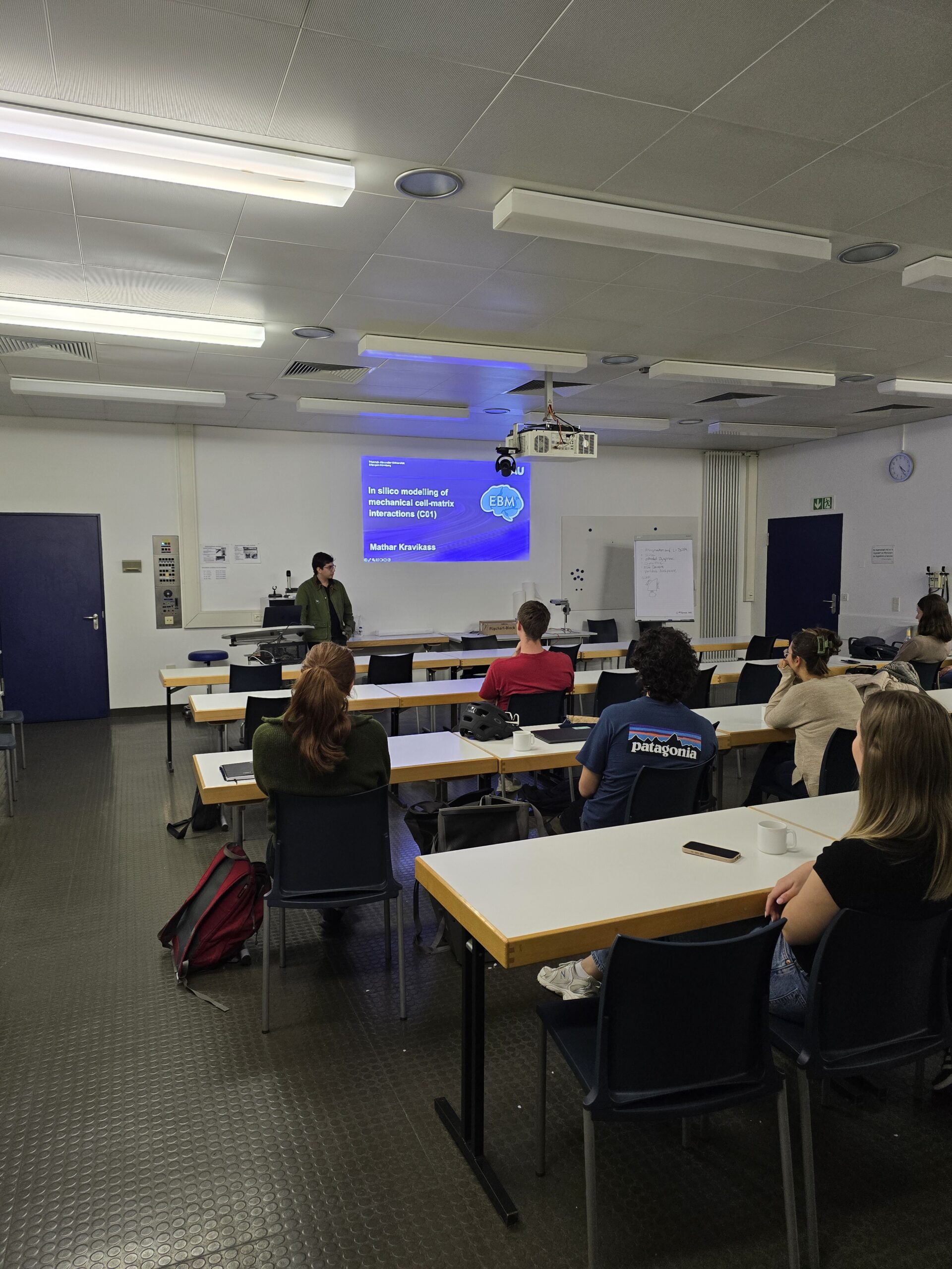
Doctoral Researchers’ Seminar by Stefan Rampp (A02) and Mathar Kravikass (C01)
The 15th EBM Doctoral Researchers’ Seminar covered a quite wide spectrum of topics: From modelling and simulation of cell aggregation phenomena to clinical aspects of brain mechanics in patients with cortical malformations.
On September 18th, Stefan Rampp (project A02: Quantitative characterization of brain malformations) and Mathar Kravikass (project C01: Continuum modeling and simulation of cell aggregation phenomena) gave updates on their respective projects.
In his project, Stefan utilizes neuroimaging methods to characterize cortical malformations and aims to bridge the gap to mechanical properties of brain tissues derived from surgical specimen and body donors. First, he provided an overview of the challenges that clinical practice faces in identifying subtle malformations, as well as the significant role that lesion identification plays in successful epilepsy surgery. He then illustrated the progress of relating diffusion-weighted imaging (DWI) to the mechanical properties of brain tissue, as determined by ex vivo testing. The second half of his presentation focused on the association of DWI with functional connectivity. He concluded by presenting two case studies that demonstrate how these relationships can be used to detect subtle malformations in clinical practice.
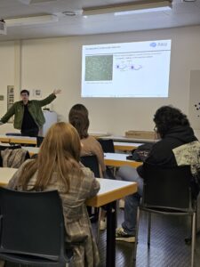
Mathar uses in-silico modeling methods to investigate the role of extracellular matrix interactions in the early development of neuronal networks. The model uses a spring bead approximation for neurites due to computational and mathematical simplicity, which allows the simulations to be scaled to a large number of neurites. In addition to the neurons, the extracellular matrix is additionally modeled as beads in two different schemes. The first being a lattice approximation, in which the beads are attached with springs, meant to emulate viscoelastic solid behavior. The second scheme is that of randomly placed beads, an approximation closer to that of a liquid. The neurites then attach to the extracellular matrix with an adhesion mechanism that temporarily links neurites to extracellular matrix beads with an additional spring force.
While using the two extracellular matrices yielded different growth patterns given the same neuron parameters, changes in the same parameters were reflected in the same way for both lattice types.
Additionally, Mathar demonstrated how this model can be expanded into synapse formation, where now intersecting neurites are allowed to form synapses, which cause the two neurons to retract to each other under the tension of the springs. In this simulation scheme, the neurons were shown to aggregate into separate clusters, connected to each other by long-reaching synapses.
Stefan Rampp, A02 & Mathar Kravikass, C01