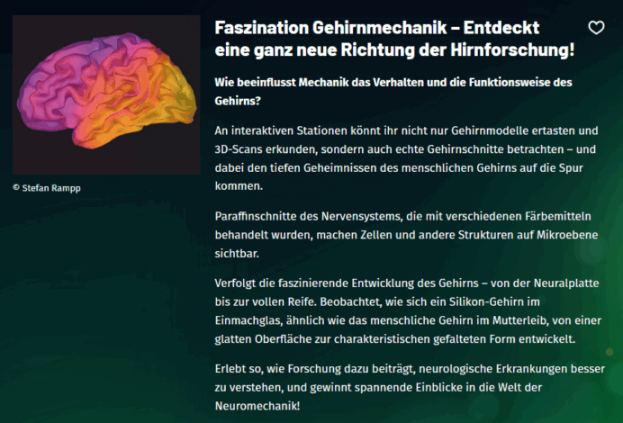
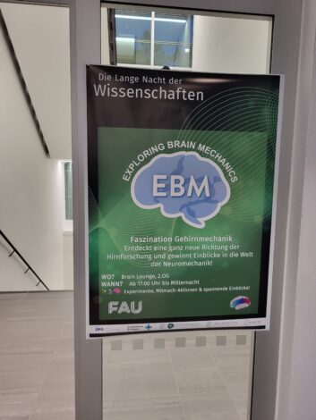
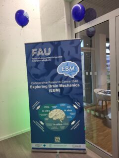
Fascination Brain Mechanics – EBM at the Long Night of the Sciences 2025
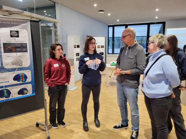
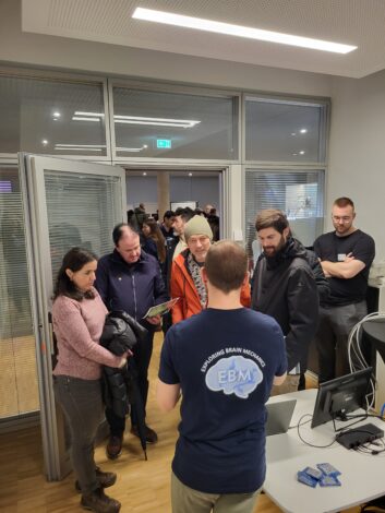
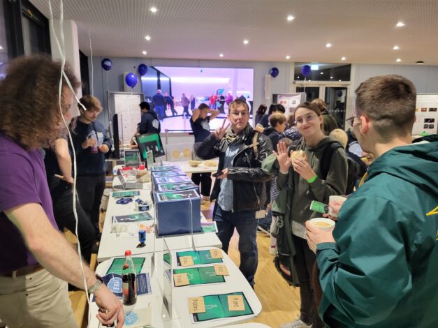
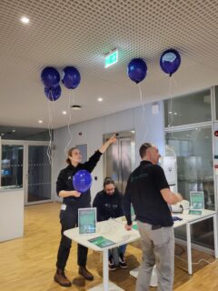
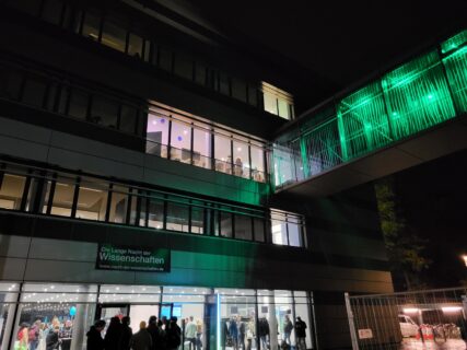
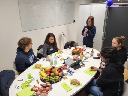
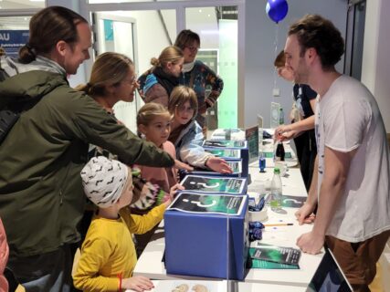
On October 25, 2025, the Collaborative Research Center 1540 “Exploring Brain Mechanics” (EBM) together with the BRAINIACS group opened its doors to the public during the Long Night of the Sciences in the Erlangen–Fürth–Nürnberg region. Under the motto “Fascination Brain Mechanics”, EBM invited visitors to the Brain Lounge at the Max Planck Center for Physics and Medicine (MPZPM) to explore the fascinating world of brain mechanics through interactive exhibits and live demonstrations.
Throughout the evening, visitors discovered how researchers at EBM study the brain at the intersection of mechanics, biology, medicine, biophysics, and material science — revealing new insights into how this complex organ works.
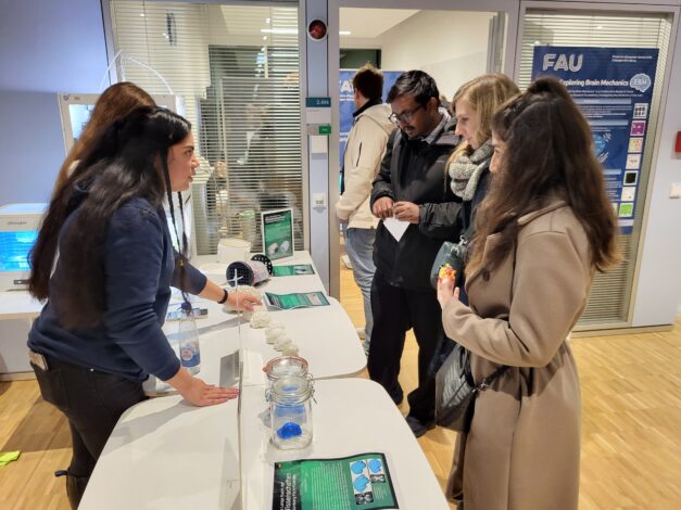
Interactive Stations to Explore and Experience
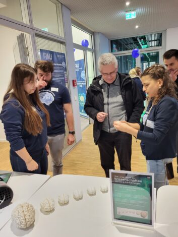
Whose brain weighs the most?
This station compared the brains of different species, showing that intelligence depends not on brain size alone but on complexity and the brain-to-body ratio. From the tiny lion brain to the massive sperm whale brain, visitors learned that the human brain is uniquely efficient and complex.
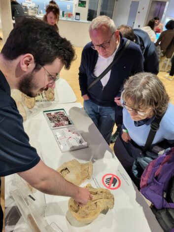
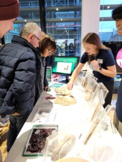
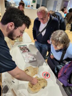
Can you guess where we hid the brain?
Many visitors tried their luck guessing what real brain tissue feels like — safely sealed and hygienically handled, of course. Pig brains were used to illustrate that brain tissue is among the softest in the human body. This hands-on activity linked directly to EBM’s research on the mechanical properties of brain tissue.
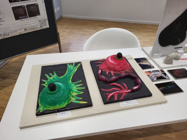
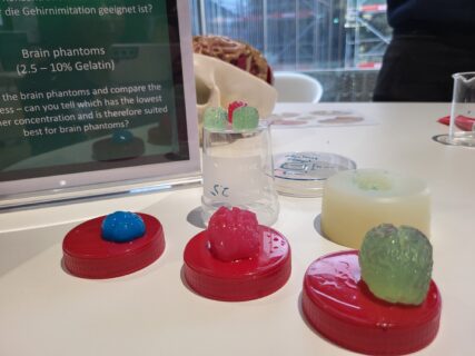
Creating artificial brain tissue
Researchers demonstrated how soft tissues can be mimicked using hydrogels — polymer networks that imitate the extracellular matrix. By varying the gel composition and stiffness, the team creates realistic brain phantoms used in experiments. Visitors were invited to touch different gelatin samples and identify which one best resembled real brain softness.
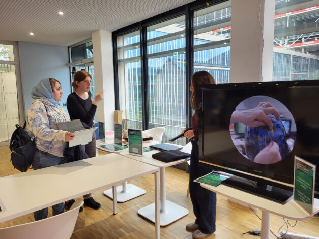
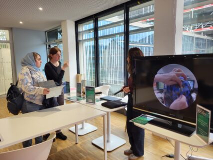
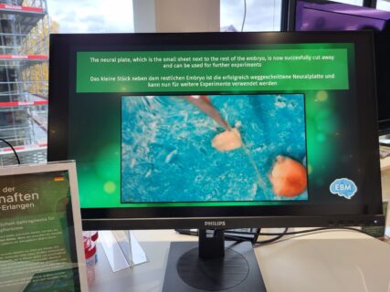
From the neural plate to the brain
Did you know that the brain once started out as a flat sheet of cells? This station showed how the neural plate folds into the neural tube and eventually forms the brain and spinal cord. A video presentation illustrated the remarkable developmental process, highlighting similarities between early human and frog brain formation.
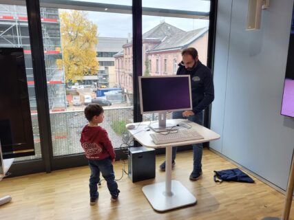
How the brain folds
Using 3D-printed models of developing brains, visitors could trace how folds and grooves emerge over gestation. An interactive computer model allowed them to experiment with mechanical parameters influencing brain folding — and observe how changes alter the resulting patterns.
Swelling brains
To understand the physical processes behind brain folding, EBM researchers use swelling polymer models that mimic the cortex and subcortex. These experiments show how mechanical instabilities can naturally lead to the complex folded structure of the human brain.
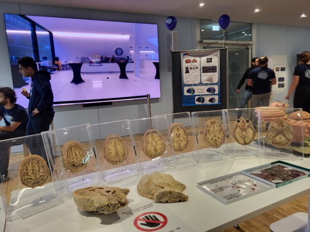
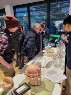
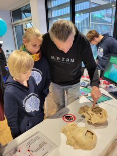
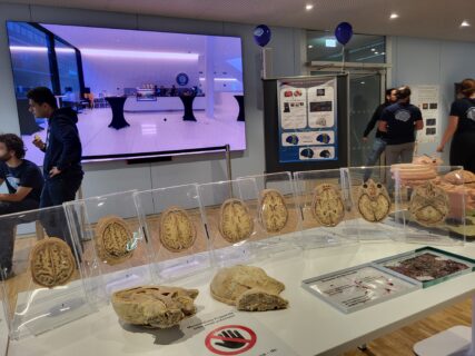
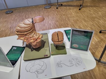
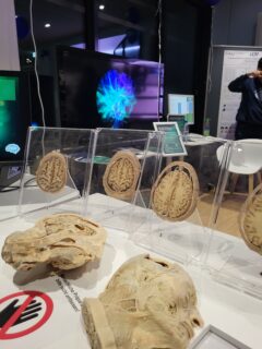
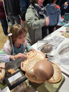
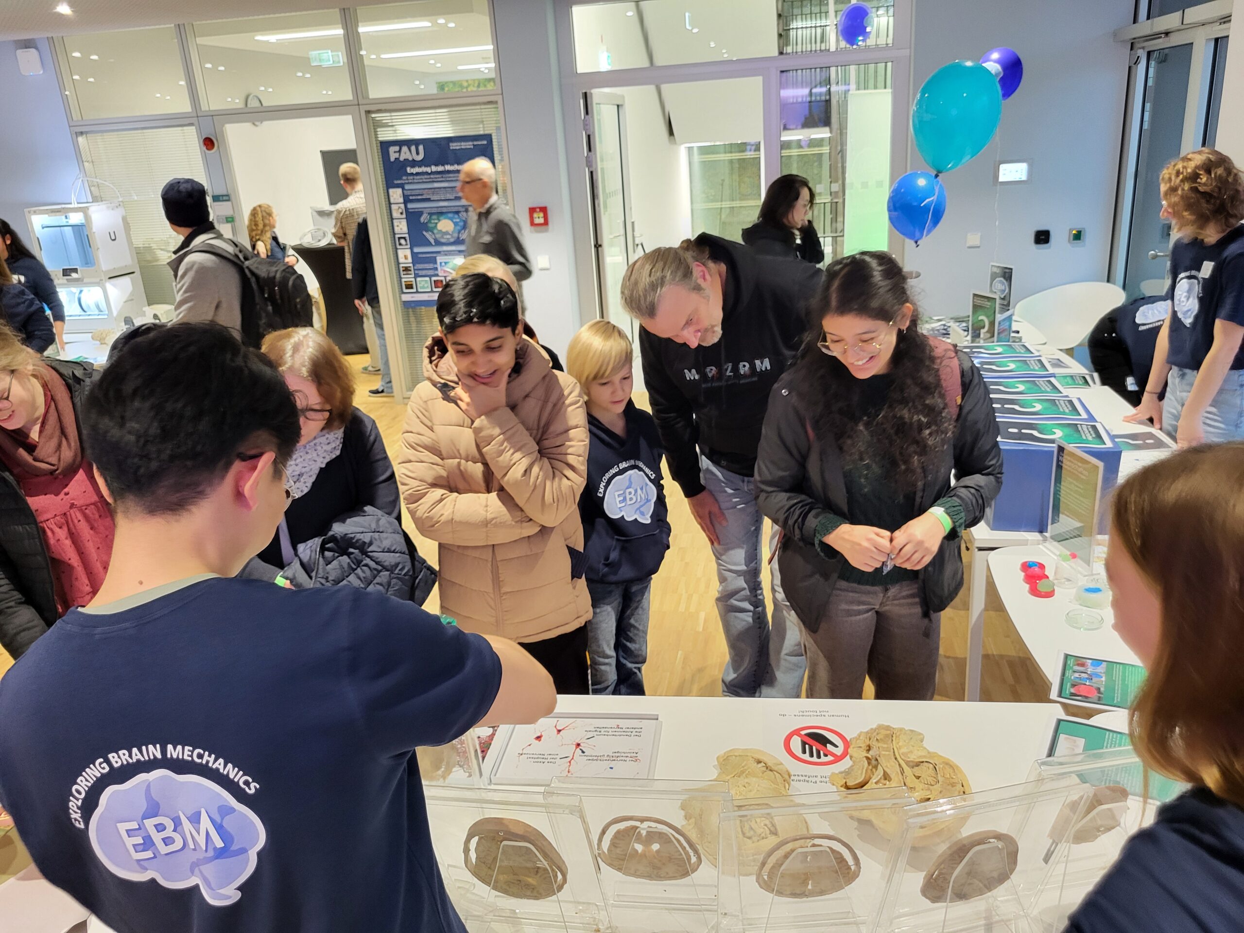
3D model of the human brain
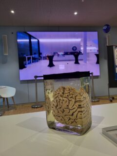
At this station, visitors explored not only detailed 3D-printed brain models but also horizontal sections of a real human brain and several preserved human brains. This rare opportunity offered a tangible, up-close encounter with the true complexity and anatomy of the human brain.
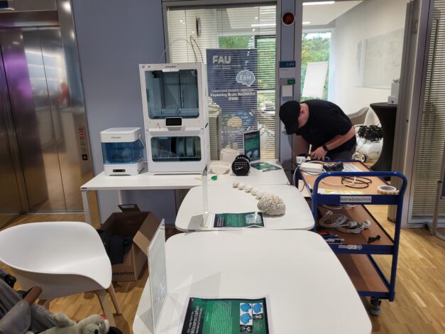
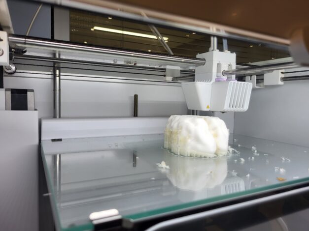
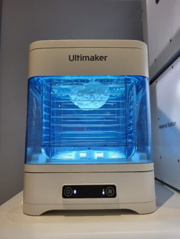
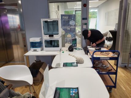
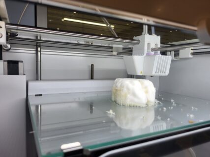
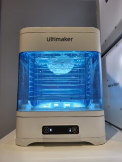
3D printing in brain research
Here, the team demonstrated how 3D printing — specifically the FDM (fused deposition modeling) method — enables the fabrication of customized holders for brain imaging or anatomical models for education and research. The exhibit included models showing brain development across different stages of pregnancy.
Magnetic Resonance Elastography (MRE)
Visitors discovered how researchers can visualize the brain’s mechanical properties without touching it. MRE combines magnetic resonance imaging (MRI) with elastography to map tissue stiffness by tracking the propagation of gentle vibrations. The displayed images illustrated how EBM uses this advanced technique to study the brain’s mechanical behavior under both healthy and pathological conditions.
Science meets organ music
A video installation featured excerpts from the film concert “The Brain – Musical Explorations”, premiered in 2024 at St. Matthäus Church in Erlangen. The projection combined organ music by Bach, Pärt, Gubaidulina, and Glass with stunning 3D visualizations from EBM’s 7-Tesla neuro-MRI research, offering a mesmerizing artistic and scientific experience.
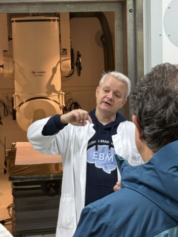
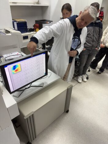
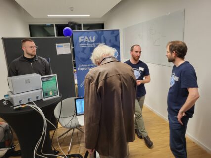
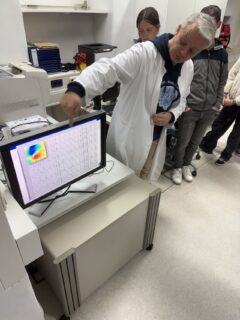
The MEG Laboratory of Neurosurgery
Right next to the MPZPM, EBM member PD Dr. med. Stefan Rampp guided visitors through the Magnetoencephalography (MEG) laboratory of the Department of Neurosurgery in six informative tours. More than 100 participants took the rare opportunity to see how MEG detects tiny magnetic fields in the brain to locate epileptic activity and study networks involved in speech, motor control, and sensory processing. The tours included a live demonstration of the MEG device’s extraordinary sensitivity and an information booth presenting ongoing research and clinical applications.
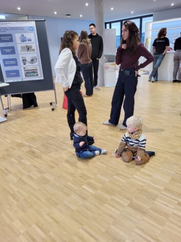
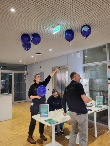
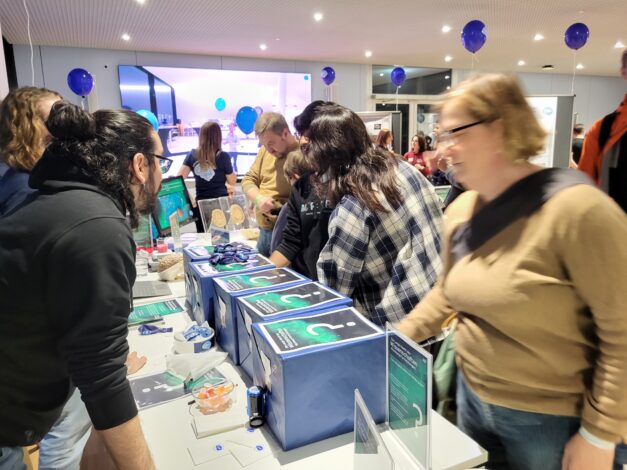
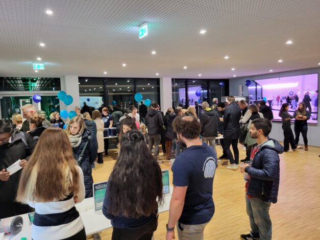
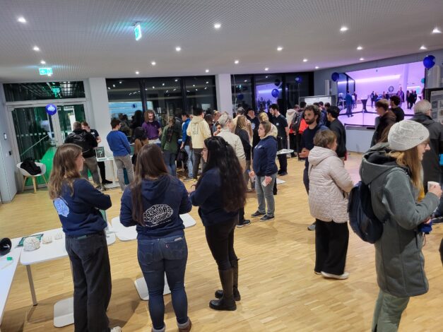
A Night of Curiosity and Inspiration
The presentation of the CRC 1540 Exploring Brain Mechanics drew considerable attention throughout the night. With creativity, enthusiasm, and hands-on demonstrations, the EBM (post)doctoral researchers succeeded in communicating the complexity and beauty of brain mechanics to a broad audience — sparking fascination and curiosity for one of the most exciting frontiers in modern science.